Related products


L-Glutamate Antibody – Rabbit Polyclonal
Validated for IF in primary neurons & brain tissues. Cited in literature
€ 449
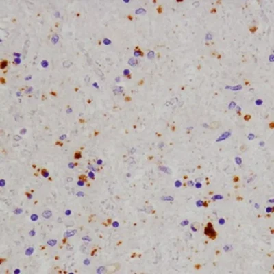
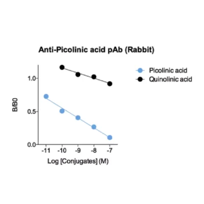
Quinolinic acid Antibody – Rabbit Polyclonal
Validated for IHC in human brain & lung FFPE samples, Cited in 4 papers
€ 449

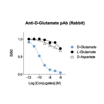
D-Glutamate Antibody – Rabbit Polyclonal
Only anti-D-glutamate antibody, Validated for IF in mouse primary neurons.
€ 449


GABA Antibody – Rabbit Polyclonal
Used for IF in primary & iPSC-derived neurons, and brain tissues. Cited in literature
€ 449


STAINperfect™ – Trial pack
Try the STAINperfect kit with 2 antibodies among 30 (neurotransmitters, amino acids, metabolites...).
Original price was: € 748.€ 397Current price is: € 397.

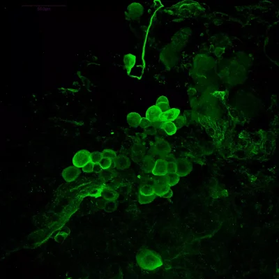
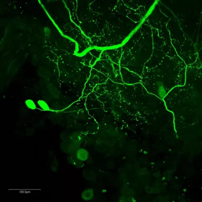
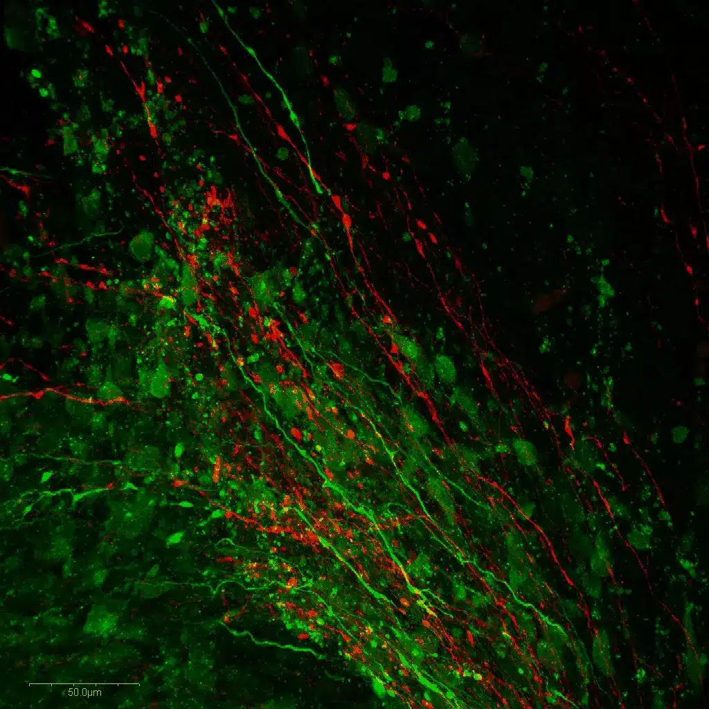
 Dopaminergic and serotoninergic neurons in crayfish brainImmunostaining of dopaminergic and serotoninergic brain neurons of a crayfish. Staining was performed with STAINperfect immunostaining kit A, following the protocol for whole mount samples. Fluorescent secondary antibodies were used and pictures were acquired by confocal imaging with high magnification.
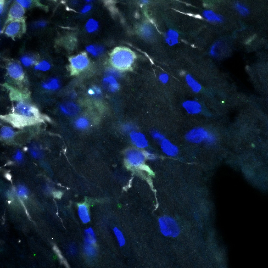
 Dopamine (green), TH (white), Hoechst (blue) in 30-day brain organoid (hiPSC)Courtesy Sarah Nickels from the Schwamborn lab
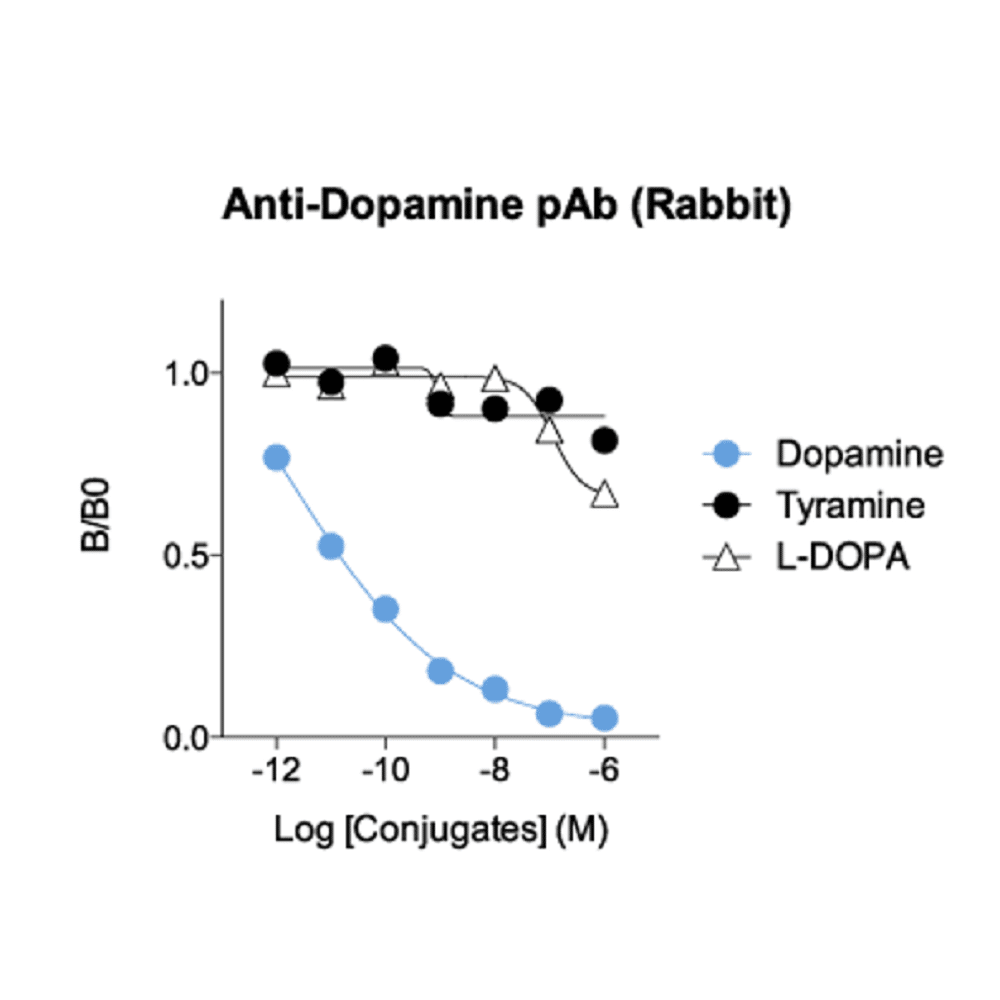
 Dopaminergic neurons of the Substantia Nigra pars compacta (SNc) in coronal rat brain sectionsRevealed by anti-dopamine (DA, rabbit polyclonal antibody #IS1005) and Tyrosine Hydroxylase (TH, #MAB5280, reference catecholaminergic neuron immunostaining). As highlighted in the overlay, DA immunoreactivity well correlates with TH immunoreactivity in the SNc, thus showing that our antibody against the DA neurotransmitter - used with the STAINperfect immunostaining kit A – is a validated tool to directly highlight DAergic systems rather than through biosynthesis enzyme immunostaining, expressed in all catecholaminergic cell populations.
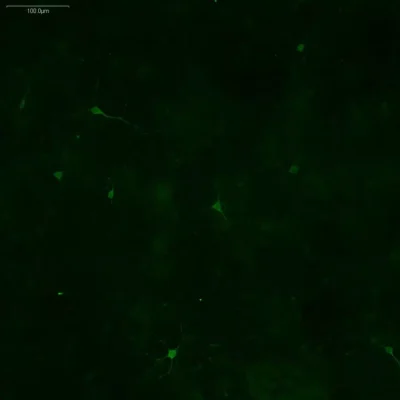
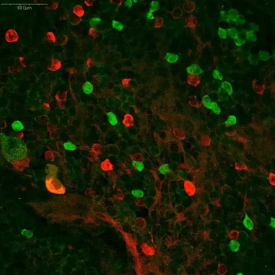
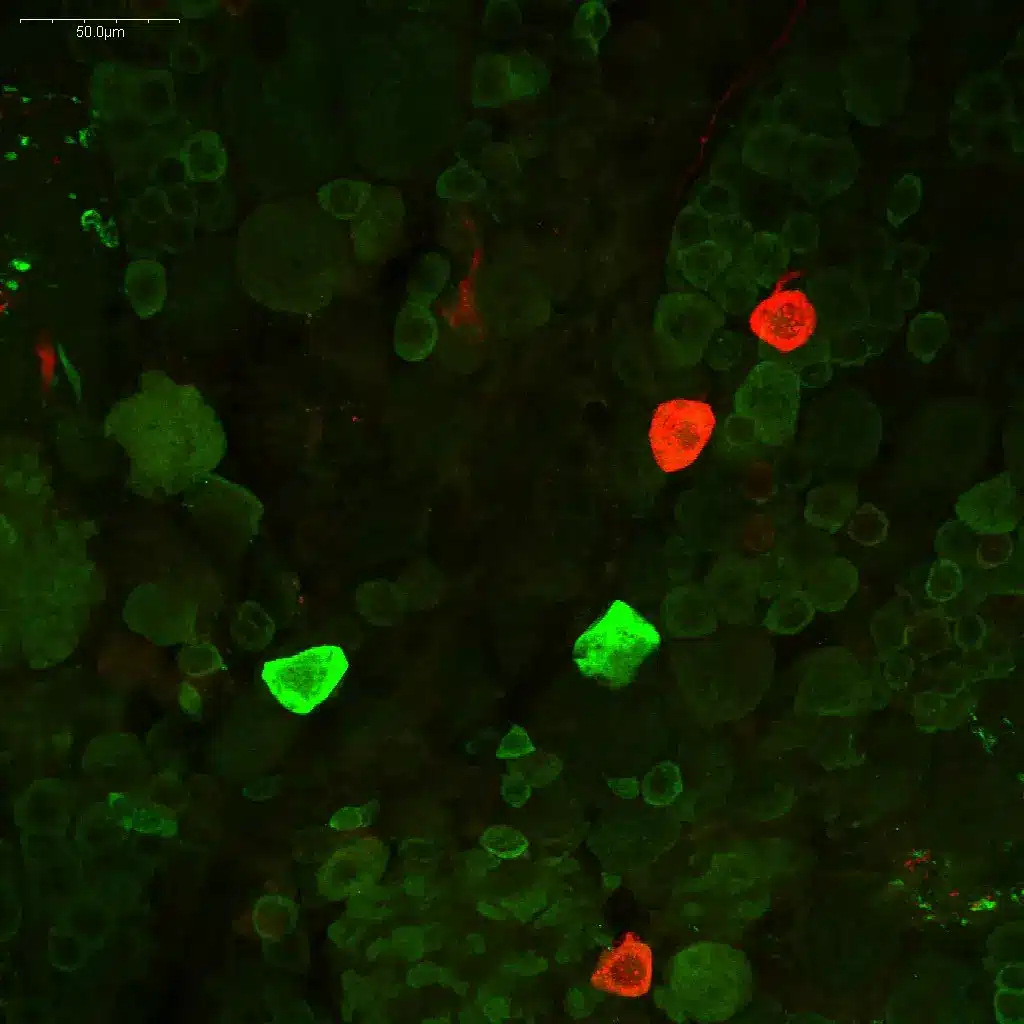
 Dopamine and Serotonin immunostaining in the CNS of embryonic mouseUsing STAINperfect immunostaining kit A, Dopamine (green) and Serotonin (red) were stained in the CNS of embryonic mouse E13.5. Staining were obtained following the protocol optimized for whole mount using IS1005 rabbit polyclonal antibody against Dopamine and IS0135 goat polyclonal antibody against Serotonin. Fluorescent conjugated secondary antibodies were then used and images captured by confocal microscopy at high magnification.
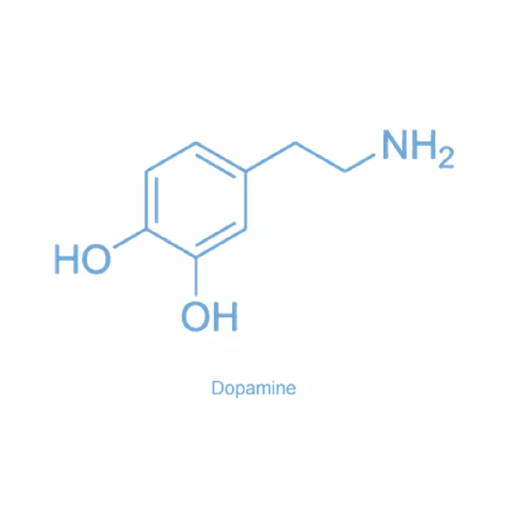
 Dopamine (3,4-dihydroxyphenethylamine)Dopamine (DA) is a catecholamine neurotransmitter and hormone synthesized from L-DOPA. Playing a key role in motor, motivational and cognitive functions, brain dopaminergic systems are found to be altered in a number of pathological states, including Parkinson's disease, attention deficit hyperactivity disorder (ADHD), drug addictions, pain and behavioral disorders. At the peripheral level, dopamine is an important regulator of blood flow, which also exerts paracrine and exocrine effects on immune cells, renal proximal tubular cells and as well as pancreatic cells.
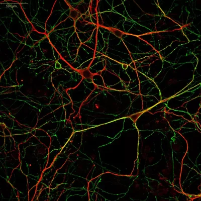
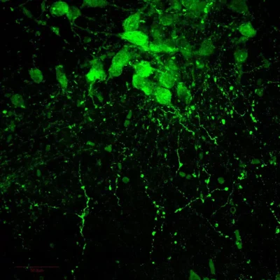
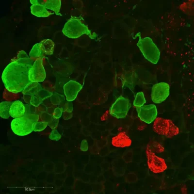
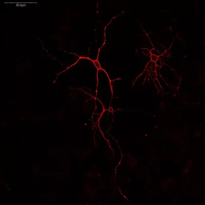
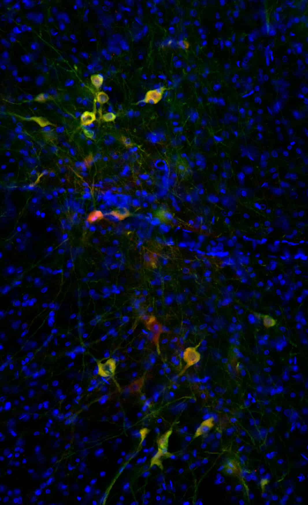
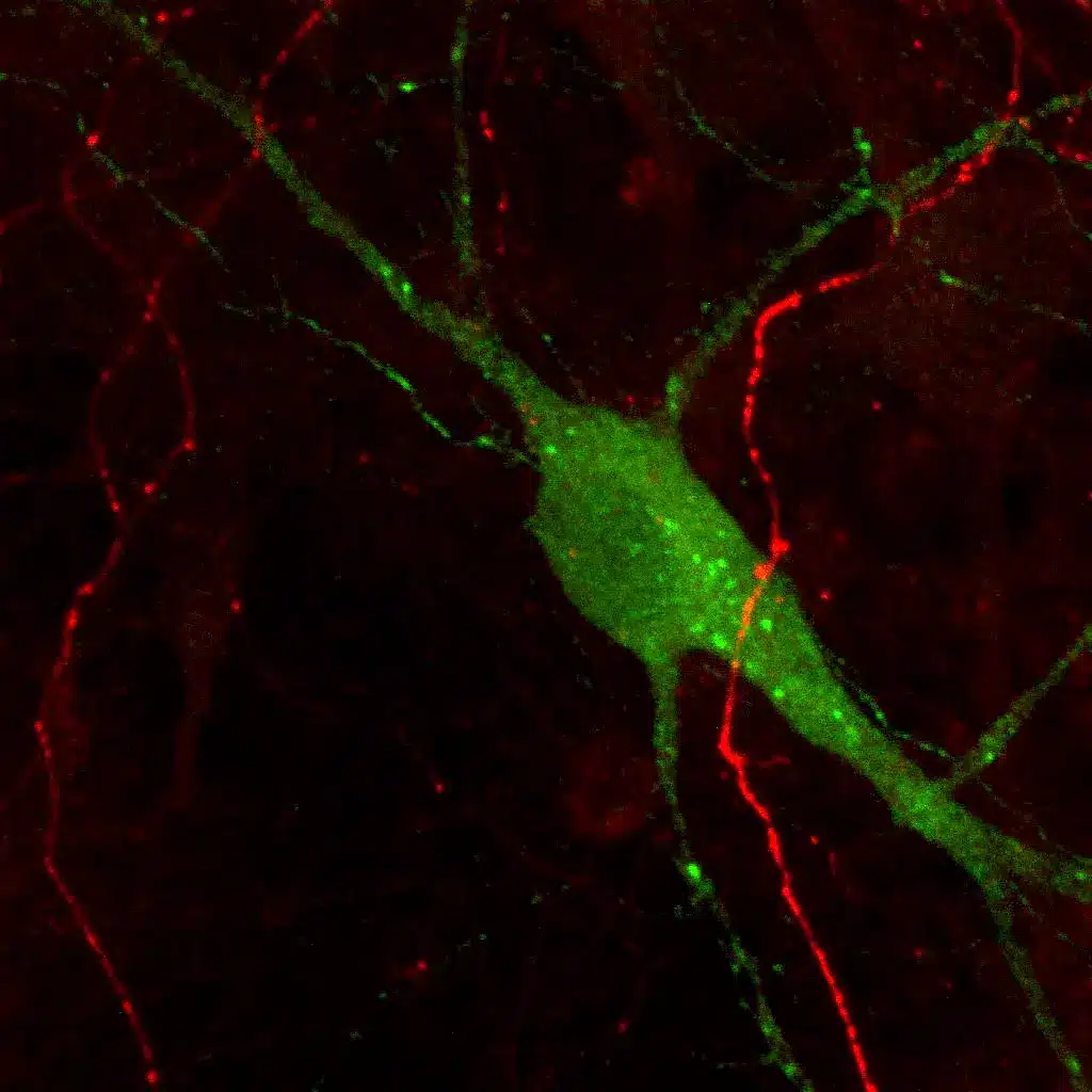
 Immunostaining of Dopamine and Serotonin in mouse culture of primary midbrain neuronsDopamine and Serotonin were stained in mouse culture of primary midbrain neurons using STAINperfect immunostaining kit A. Staining were obtained following the protocol optimized for cell culture using IS1005 rabbit polyclonal antibody against Dopamine and IS0135 goat polyclonal antibody against Serotonin. Fluorescent conjugated secondary antibodies were then used and imaged by confocal imaging.


 and Tyrosine Hydroxylase (TH, #MAB5280, reference catecholaminergic neuron immunostaining). As highlighted in the overlay, DA immunoreactivity well correlates with TH immunoreactivity in the SNc, thus showing that our antibody against the DA neurotransmitter - used with the STAINperfect immunostaining kit A – is a validated tool to directly highlight DAergic systems rather than through biosynthesis enzyme immunostaining, expressed in all catecholaminergic cell populations.")
 and Serotonin (red) were stained in the CNS of embryonic mouse E13.5. Staining were obtained following the protocol optimized for whole mount using IS1005 rabbit polyclonal antibody against Dopamine and IS0135 goat polyclonal antibody against Serotonin. Fluorescent conjugated secondary antibodies were then used and images captured by confocal microscopy at high magnification.")

 is a catecholamine neurotransmitter and hormone synthesized from L-DOPA. Playing a key role in motor, motivational and cognitive functions, brain dopaminergic systems are found to be altered in a number of pathological states, including Parkinson's disease, attention deficit hyperactivity disorder (ADHD), drug addictions, pain and behavioral disorders. At the peripheral level, dopamine is an important regulator of blood flow, which also exerts paracrine and exocrine effects on immune cells, renal proximal tubular cells and as well as pancreatic cells.")

You may also like…
STAINperfect™ immunostaining kit A
Compatible with 30+ antibodies. Cell culture/Whole mounts/Tissue sections. Cited in 20+ papers
€ 299€ 349


GABA Antibody Chicken Polyclonal
Validated for IF in primary & iPS-derived neurons, and brain tissues. Cited in literature
€ 449
Glutamate Antibody (L) – Mouse Monoclonal
Validated for IF in primary neurons & iPSC-derived neurons. Cited in literature
€ 449
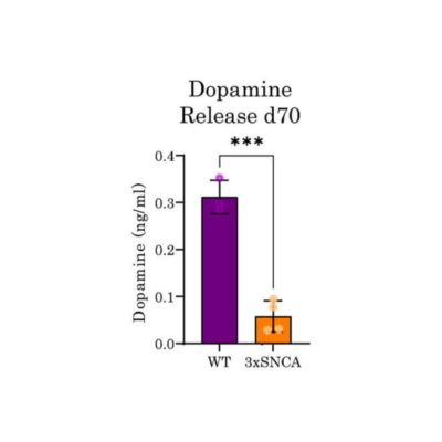
Ultra-Sensitive Dopamine ELISA kit – Any sample
Ultra-sensitive, ≥ 1µl sample, Cited in 40+ papers
€ 629
