The clear path to small molecule visualization & quantification
Latest news Scientific Communications

GABA: A Neurotransmitter Behind Immunotherapy Resistance in TLS-Positive Tumors
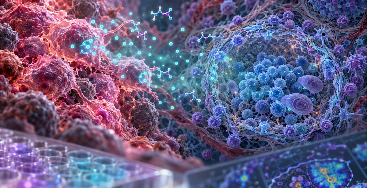
Tumor-derived GABA and resistance to immunotherapy in TLS-positive tumors

GABA: A Neurotransmitter Behind Immunotherapy Resistance in TLS-Positive Tumors
A new Cancer Cell study reveals an unexpected role for GABA in the tumor microenvironment, and highlights the need for tools that can both visualize and quantify this small immunoregulatory metabolite.

Gamma-aminobutyric acid (GABA) is best known as the major inhibitory neurotransmitter of the central nervous system. A growing body of research shows that its biology extends well beyond neuronal signaling.

A study published in Cancer Cell in 2026, “GABA promotes resistance to immunotherapy in patients with TLS-positive tumors”, identifies tumor-derived GABA as a potential mediator of resistance to immune checkpoint inhibitors (ICIs) in tumors containing tertiary lymphoid structures (TLSs).

The findings extend GABA from neuroscience into tumor immunology and cancer metabolism. They also show why researchers need reliable ways to detect GABA directly in tissues, cells and biological fluids.

Tumor-derived GABA is associated with resistance to immunotherapy

Tertiary lymphoid structures are organized immune-cell aggregates that develop within or around tumors. They have been associated with favorable responses to ICIs in several cancer types.

Yet many patients with TLS-positive tumors do not respond. To understand why, Hernández-Verdin and colleagues, working with teams at the Centre de Recherche des Cordeliers, Institut Bergonié and Gustave Roussy, analyzed clear-cell renal cell carcinoma (ccRCC) and soft-tissue sarcoma (STS) treated with immune checkpoint inhibitors. ImmuSmol and Explicyte scientists Alban Bessède and Jean-Philippe Guégan are co-authors of the study.

In 58 TLS-positive ccRCC tumors from the BIONIKK trial, GABA-related gene signatures were enriched in non-responders. 85% of patients with a high GABA signature did not respond, compared with 26% of those with a low signature. The association was supported in additional ccRCC cohorts (NIVOREN, IMmotion150/151) and in TLS-positive sarcomas from the PEMBROSARC trial. It was seen only in TLS-positive tumors treated with immunotherapy, not in TLS-negative tumors or in patients treated with tyrosine kinase inhibitors.

In ccRCC, GABA production was mainly associated with a population of malignant cells with a proximal-tubule-like phenotype. Nerve-related signatures and nerve density did not explain the association. TLSs located close to these GABA-producing tumor cells showed signs of impaired immune function, including altered B-cell maturation, reduced IgG expression, impaired antigen presentation and increased GABA-receptor expression.

The study therefore suggests that the presence of a TLS is not enough on its own. The metabolic environment around it matters too.

GABA directly affects B-cell function

The investigators then asked whether GABA itself could act on immune cells. In activated human B cells from healthy donors, GABA reduced HLA-DR expression, proliferation and immunoglobulin secretion. The reduction in HLA-DR did not depend on GABA receptors, whereas the effect on B-cell growth was rescued by blocking the GABAB receptor. This points to both metabolic and receptor-mediated mechanisms.

The pathway was also tested in vivo. In a TLS-positive mouse sarcoma model, the GABA-synthesis inhibitor 3-MPA had no effect on its own but enhanced the antitumor activity of anti-PD-1. Compared with anti-PD-1 alone, the combination increased B-cell density and proliferation inside TLSs, with a trend toward more plasma cells.

Together, these data position GABA as an immunoregulatory metabolite in the tumor microenvironment, not only a neurotransmitter. The authors note that confirmation in patient-derived immune cells and further clinical cohorts will be needed.

Studying GABA requires both spatial and quantitative approaches

The study also measured GABA directly. Intratumoral GABA concentrations, determined by mass spectrometry, correlated inversely with B-cell density inside TLSs. Spatial metabolomics mapped GABA-rich regions around TLSs.

These analyses were combined with transcriptomics and immunophenotyping, which illustrates an important point for GABA research. Expression of GAD1/GAD2, GABA receptors or GABA-related gene signatures describes the pathway, but it does not answer two basic questions:

  • Where is GABA itself located?
  • How much GABA is present in the sample?

Direct GABA detection complements transcriptomic and enzyme-based approaches. This is where antibody-based imaging and targeted quantitative assays come in.


Direct visualization of GABA with ImmuSmol anti-GABA antibodies

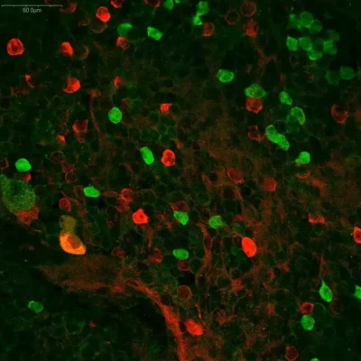
The Cancer Cell study lists the ImmuSmol rabbit polyclonal anti-GABA antibody IS1006 among the reagents in its Key Resources Table.

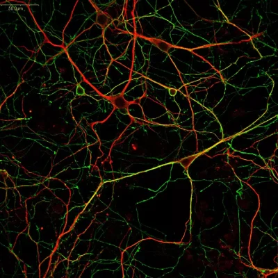
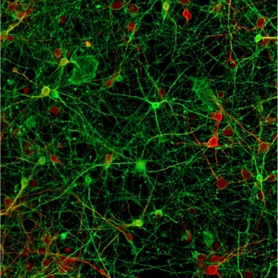
ImmuSmol provides anti-GABA antibodies raised in rabbit, mouse and chicken, which gives researchers flexibility for multiplex immunostaining. The portfolio was developed to visualize GABA directly in cell cultures, whole mounts and tissue sections.

These antibodies have a published track record in several experimental systems. The chicken polyclonal IS1036, cited in five publications, has been used in mouse primary neurons, crayfish tissues and rodent brain tissue. Its chicken host makes it easy to combine with rabbit or mouse antibodies in multiplex panels. The rabbit polyclonal IS1006 has been used in human stem-cell-derived neurons and rabbit olfactory bulb tissue. The mouse monoclonal IS039 has been used in rat primary neurons and human iPSC-derived midbrain organoids. All three are optimized for use with the STAINperfect immunostaining kit A, which is designed for small-molecule detection.

As GABA biology expands into cancer immunology, finding the cellular and spatial sources of the metabolite can matter as much as measuring its overall abundance.

Explore ImmuSmol anti-GABA antibodies →


Quantifying GABA across biological matrices
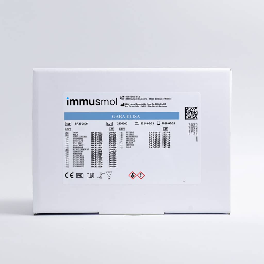
GABA ELISA kit BA-E-2500Imaging answers “where?”. Quantitative assays answer “how much?”.

The ImmuSmol GABA ELISA kit BA E-2500 measures GABA quantitatively in biological samples. It uses a 96-well format and requires approximately 100–300 µL of sample depending on the matrix. It works with samples from any species, with measurement ranges of 25–2,500 or 75–7,500 ng/mL depending on the assay configuration.

A key feature of the assay is that it is not restricted to serum or plasma. It has been used in the scientific literature across a wide range of biological materials:

Biological materialExample of published use
Brain tissueGABA quantified in rodent brain regions in neuropharmacology studies
Spinal cord tissueGABA measured in spinal-cord protein extracts in a study of the astrocytic MAOB–GABA axis after spinal-cord injury
Zebrafish tissueGABA quantified in homogenized larval zebrafish heads in an Epilepsia study of KCNB1 loss of function
Cell-culture mediaGABA released into culture media by human iPSC-derived GABAergic neurons
Extracellular vesiclesGABA loading into extracellular vesicles, reported in Stem Cell Research & Therapy
Airway epithelial cellsSmall airway epithelial cells exposed to estrogen and a tobacco carcinogen (Al-Wadei et al., Lung Cancer, 2009)

With 15+ publications, this range of matrices matters for translational studies, where the same metabolite may need to be measured at several levels: tumor tissue, experimental models, cell-culture supernatants and circulating samples.

Explore the GABA ELISA kit →

From GABA pathway signatures to direct metabolite measurement

The Cancer Cell study is an important example of how a metabolic pathway can shape antitumor immunity. It also shows the value of a multimodal strategy.

Transcriptomics can identify GABA synthesis and receptor programs. Mass spectrometry provides detailed metabolic and spatial profiling. Immunostaining can show where GABA accumulates within tissues. A targeted ELISA offers a practical way to quantify it across larger experimental series and multiple biological matrices.

These approaches complement each other rather than compete. The ImmuSmol portfolio covers both dimensions of GABA analysis:

  • Visualize GABA with anti-GABA antibodies used in cellular, tissue and organoid models.
  • Quantify GABA with a literature-supported ELISA applicable to a broad range of biological matrices.

As research reveals more roles for GABA outside the nervous system, including in the regulation of antitumor immunity, direct and flexible ways to detect it are likely to become more valuable in both preclinical and translational research.

Reference: Hernández-Verdin I. et al. GABA promotes resistance to immunotherapy in patients with TLS-positive tumors. Cancer Cell 44 (2026). doi:10.1016/j.ccell.2026.06.006

For research use only.

Search
×
0
0
Your Cart
Your cart is emptyReturn to Shop