Validated monoclonal anti-GABA antibody to evaluate GABAergic system network/function by immunofluorescence
June 13, 2019 2025-06-10 16:51Validated monoclonal anti-GABA antibody to evaluate GABAergic system network/function by immunofluorescence
Validated monoclonal anti-GABA antibody to evaluate GABAergic system network/function by immunofluorescence
Gamma-AminoButyric Acid (GABA) is the major inhibitory neurotransmitter in the developmentally mature vertebrate central nervous system, which binds GABAA and GABAB receptors thereby causing hyperpolarization and neuronal activity inhibition.
GABA system/function alterations are known to be associated with many psychiatric and neurological disorders, e.g. anxiety, depression, schizophrenia, sleep disorders, insomnia…
The 7F5.2B4.1B4 monoclonal antibody has been developed and validated to directly react with and label GABA, thereby making it a suitable tool to evaluate GABAergic system networks / function by immunofluorescence (IF) and immunohistochemistry (IHC), on varied types of biological materials.

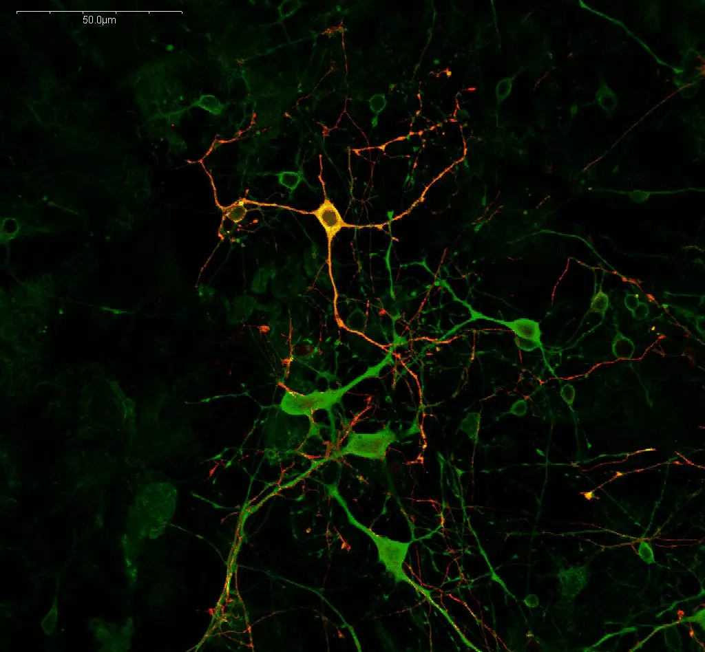
GABAergic neuronal network in a mature rat cortical culture revealed through GABA immunoreactivity detection using either our mouse monoclonal GABA antibody (#IS039) in green or rabbit polyclonal GABA antibody (#IS1006) in red, with the STAINperfect immunostaining kit A. GABAergic network is identically revealed by both antibodies (yellow on the overlay) which evidence the cortical GABAergic neurons as well as their rich and dense neuritic extensions.

GABAergic network and glutamatergic cell populations in a mature rat cortical culture revealed by GABA (mouse monoclonal antibody #IS039) and L-glutamate (rabbit polyclonal antibody #IS1001) immunoreactivity. As expected, our antibodies, appropriately used with the STAINperfect immunostaining kit A, highlight the rich cortical GABAergic system (cell bodies and branches – displaying a punctiform staining) as well as the glutamate-positive cell population. While GABA neurons are also glutamate-positive (some are pointed by the arrows), arrowheads point glutamate-positive GABA-negative cells, which can be either astrocytes or glutamatergic neurons.
