No products in the cart.
Dopamine Antibody – Rabbit Polyclonal
Ref: IS1005CatecholaminesPolyclonal AntibodiesICCIFIHCNeuromediators
") +
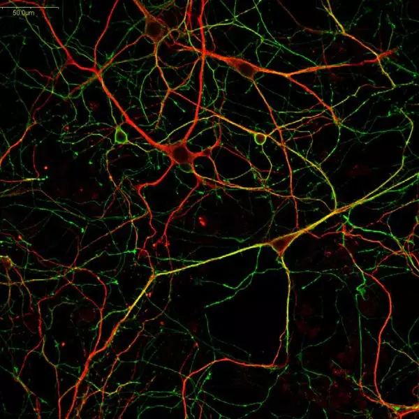
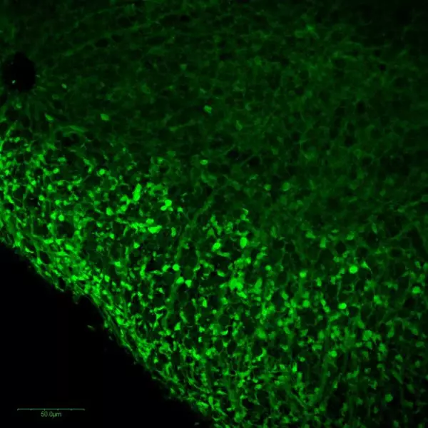
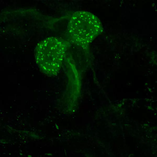
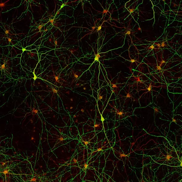
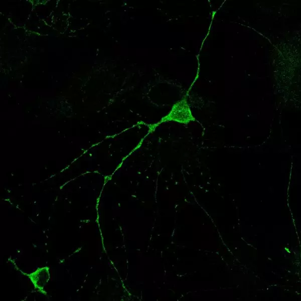
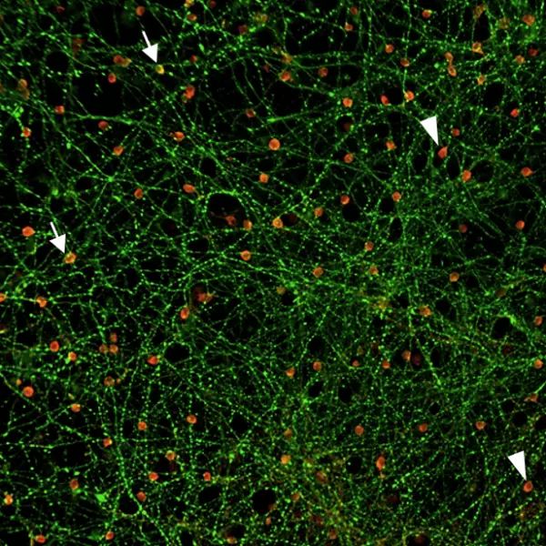
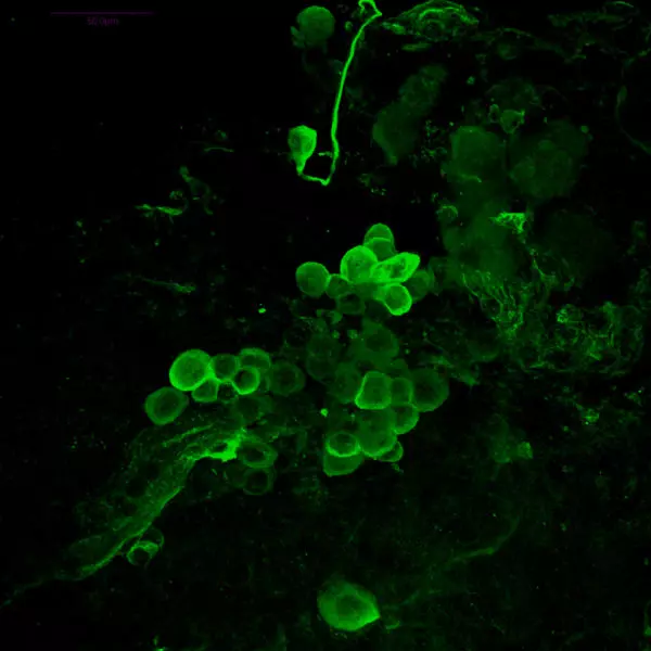
+Dopamine imaging in mouse embryonic mesencephalic neurons (E13.5)
Using STAINperfect immunostaining kit A, Dopamine (green) was detected within mouse embryonic mesencephalic neurons (E13.5) following the protocol optimized for whole mount samples. Stainings were performed using ImmuSmol rabbit anti-Dopamine polyclonal Ab (IS1005) and standard mouse anti-TH antibody. Fluorescent conjugated antibodies were used and images acquired by confocal imaging.


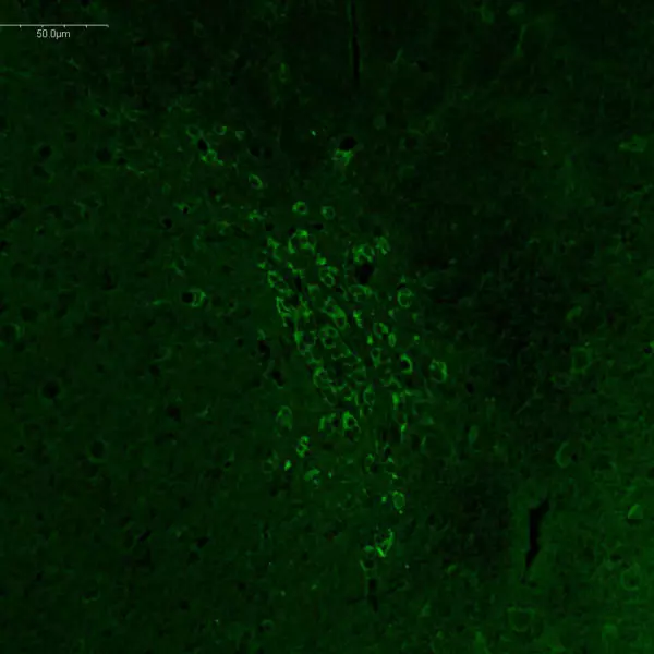
 in coronal rat brain sections")

")
DatasheetMSDS
Cited in 11 papers, the anti-dopamine antibody IS1005 was used to evidence dopamine in mouse primary & iPS-derived neurons, human brain tissues and stem-cell derived brain organoids. Combined with the STAINperfect immunostaining kit A, this rabbit polyclonal antibody enables high-quality dopamine imaging.
| Clonality | Polyclonal antibody |
| Host | Rabbit |
| Reactivity | Reacts with all species |
| Tested samples | Whole mounts, cell culture, tissue sections, organoids |
| Staining procedure Format | STAINperfect immunostaining kit A |
| Format | 50µl (approx. 40 tissue sections) |
| References | Cited in 11 papers |
They published with this product see papers


Product overview
| Product name | Dopamine antibody – Rabbit pAb |
| Synonyms | Anti-Dopamine antibody Anti-3,4-dihydroxyphenethylamine antibody Anti-DA antibody Anti-hydroxytyramine antibody Anti-oxytyramine |
| Immunogen | Conjugated Dopamine |
| Specificity | When tested in competitive ELISA, the anti-conjugated Dopamine antibody did not show any significant cross reactivity with Tyramine and L-Dopa conjugates |
| Volume | 50 µl |
| Lot number | 140301 |
| Expiration date | 2025-08-01 |
Storage
| Form | Liquid |
| Purity | Purified anti-serum |
| Storage | Store at +4°C for short term (6 months). Aliquot and store at -20°C for long term. Avoid repeated freeze / thaw cycles |
| Material safety datasheet | Download MSDS |
| IF – Cell cultures, Whole mounts, Tissue sections | Dilute antibody with the antibody diluent provided in the STAINperfect immunostaining kit A. Use at 1/250 -1/1000 dilution. Follow the STAINperfect protocol suited to your sample |
| Comments | Optimal working dilutions must be determined by the end-user |
| Restrictions | For research use only |
| Full protocol | Download STAINperfect protocol for dopamine staining |
| Protocols-at-a-glance |
 | 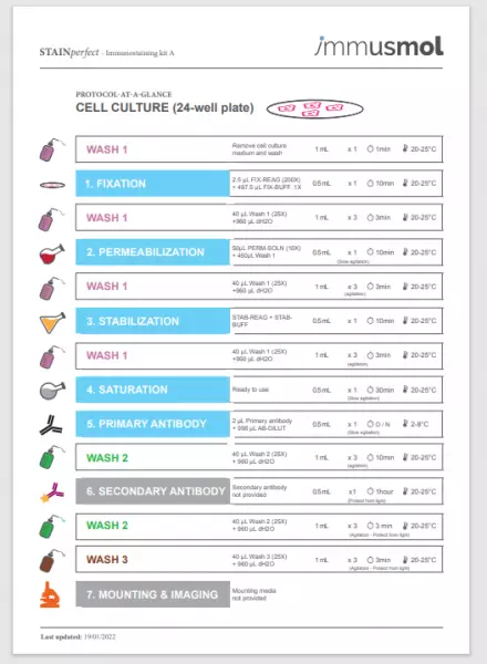 | 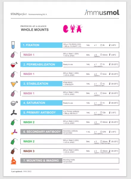 | 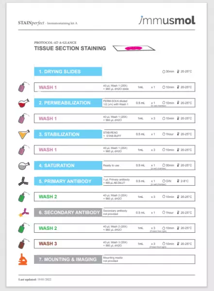 |
| Complete Instructions for Use | Protocol-at-a-glance for cell cultures | Protocol-at-a-glance for whole mounts | Protocol-at-a-glance for tissue sections |
Product citation
- Treating Parkinson’s Disease with Human Bone Marrow Mesenchymal Stem Cell Secretome: A Translational Investigation Using Human Brain Organoids and Different Routes of In Vivo Administration
Check the article
Authors : Mendes-Pinheiro et al., Cells
2023-11 - Production of Highly Uniform Midbrain Organoids from Human Pluripotent Stem Cells
Check the article
Authors : Yao et al., Stem Cells International
2023-09 - Dopamine Negatively Regulates Insulin Secretion Through Activation of D1-D2 Receptor Heteromer
Check the article
Authors : Uefune et al., Diabetes
2022-06 - Alkyne-Tagged Dopamines as Versatile Analogue Probes for Dopaminergic System Analysis
Check the article
Authors : Nuriya et al., Analytical Chemistry
2021-07 - Hair-Follicle-Associated Pluripotent (HAP) Stem Cells Can Extensively Differentiate to Tyrosine-Hydroxylase-Expressing Dopamine-Secreting Neurons
Check the article
Authors : Yamane et al., Cells
2021-04 - Single-cell transcriptomics reveals multiple neuronal cell types in human midbrain-specific organoids
Check the article
Authors : Smits et al., Cell & tissue research
2020-07 - Reproducible generation of human midbrain organoids for in vitro modeling of Parkinson’s disease
Check the article
Authors : Nickels et al., Stem Cell Research
2020-06 - Apoptosis signal regulating kinase 1 deletion mitigates α-synuclein pre-formed fibril propagation in mice
Check the article
Authors : Zhang et al., Neurobiology of aging
2019-09 - Modeling Parkinson’s disease in midbrain-like organoids
Check the article
Authors : Smits et al., NPJ Parkinson's disease
2019-04 - miR-34b/c Regulates Wnt1 and Enhances Mesencephalic Dopaminergic Neuron Differentiation
Check the article
Authors : De Gregorio et al.,
2018-03 - Derivation of Human Midbrain-Specific Organoids from Neuroepithelial Stem Cells
Check the article
Authors : Monzel et al., Step cell reports
2017-05