Generic selectors
Exact matches only
Search in title
Search in content
Post Type Selectors
Cart

No products in the cart.

Immusmol's News

03
2024

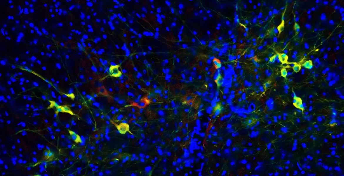
Beyond TH: Imaging Dopamine Content in Midbrain Organoids for Parkinson’s Disease Modeling

Dopamine TH staining antibody elisa organoids Parkinson's disease modeling

Since the early papers describing the generation of stem cell-derived midbrain organoids in 2016-2017, research teams have aimed to demonstrate the presence of functional dopaminergic neurons in these 3D microtissues. One widely trusted and accepted marker for dopaminergic function is tyrosine hydroxylase (TH) – the enzyme converting L-Tyrosine into L-Dopa, the precursor of Dopamine.

However, TH expression does not necessarily indicate that neurons are metabolically active and produce dopamine. Therefore, several groups interested in organoid-based modeling of Parkinson’s disease have combined standard markers of dopaminergic neurons, such as TH and GIRK2, with direct staining of dopamine using anti-hapten antibodies, as well as ELISA kits for quantifying dopamine release in supernatant.

Five selected articles demonstrating dopamine biosynthesis in human midbrain organoids

Seminal paper (>350 citations) describing the 3D differentiation of human neuroepithelial stem cells into midbrain-specific organoids containing dopamine-producing neurons at day 50 post-differentiation

In 2017, the group of Jens Schwamborn from the Luxembourg Centre for Systems Biomedicine (LCSB) released one of the first protocol to generate stem-cell derived midbrain organoids. The team demonstrated the presence of midbrain-specific dopaminergic neurons at day 50 post-differentiation, using markers such as TH, TUJ1, GIRK2, CALBINDIN, Dopamine transporter, as well as dopamine itself, using the IS1005 antibody.

Midbrain organoid whole mount stainings of TH & dopamine, and quantification of dopamine release at day 70 post-differentiation

In 2019, the Schwamborn group, together with colleagues from the Max Planck Institute and the Hannover Medical School, published an alternative protocol (>200 citations) for the generation of midbrain organoids, starting with hiPSC-derived midbrain floor plate neural progenitor cells (mfNPCs). They demonstrated the presence of DA-producing neurons after 70 days through TH & dopamine co-staining, as well as a significant increase in dopamine release over differentiation (Day 35, Day 70) measured by ELISA . They also validated this protocol for the in vitro modeling of Parkinson’s disease using iPSCs derived from Parkinsonian patients.

Optimized midbrain organoids containing functional dopaminergic neurons after 30 days (whole mount stainings), and 6-OHDA exposure to model Parkinson’s disease

In 2020, the Schwamborn group optimized their 2019 protocol to reduce batch-to-batch variability and avoid the formation of necrotic cores. They demonstrated dopaminergic functionality in resulting 3D microtissues through co-stainings for TH, GIRK2, dopamine transporter, and dopamine content.

TH, GIRK2 and dopamine in whole mount stainings of midbrain organoids at day 30 post-differentiation, prior to 6-OHDA lesion and treatment with MSC secretome

In 2023, to assess the therapeutic potential of Mesenchymal Stem Cell (MSC) secretome, a team from the University of Minho, Portugal, used the protocol described by Smits et al [2] to generate midbrain organoids recapitulating PD-like dopaminergic degeneration. To demonstrate dopaminergic function before 6-OHDA exposure, they stained organoid sections for TH, GIRK2 and Dopamine (IS1005 pAb).

A High-throughput organoid platform for drug discovery and toxicity studies: demonstrating the presence of mature dopaminergic neurons (TH, GIRK2) and the production of dopamine & neuromelanin-like granules in micro-midbrain organoids (Day 30 post-differentiation)

A public-private consortium from South Korea unveiled in 2023 a high-throughput platform for the generation of functional micro-midbrain organoids. Designed for large-scale drug discovery and toxicity studies, their platform is based on a single-step process in 96- and 384-well plates, which produces mature brain organoids with minimal variability within 4 weeks. Exhibiting cellular diversity, the resulting micro-organoids were analyzed for their dopaminergic content, using TH and GIRK2, together with the production of dopamine and neuromelanin-like granules.

How to stain midbrain organoids for dopamine? A quick practical guide:

The Schwamborn group, in their first paper, described a protocol to prepare tissue sections of midbrain organoids, using paraformaldehyde fixation and low-melting point agarose. Primary dopamine antibody IS1005, compatible with PFA-fixed tissues, was successfully used following standard protocols.

However, to minimize background noise in tissue sections and label dopamine in whole mounts, we recommend using the STAINperfect staining kit. Compatible with 10 antibodies to key neurotransmitters and designed for multiplexed imaging, this immunostaining kit was used in 2020 by the team of Jens Schwamborn and colleagues from the RIKEN & University of California to evidence the presence of neurons producing dopamine, glutamine, GABA, and serotonin in human midbrain organoids.

Learn more about anti-dopamine antibody IS1005 and research ELISA kit for the quantification of dopamine in low-volume sampes (1 µl min.)

Dopamine Antibody – Rabbit Polyclonal

IS1005

Used for IF in mouse primary & iPS-derived neurons, human organoids & brain tissues
Cited in 11 papers

From: 440.00
VIEW PRODUCTADD

Dopamine ELISA kit – Ultra-Sensitive
Any sample

BA-E-5300R

Use Research only
Samples Any type, ≥ 1µl
Key Feature High Sensitivity

465.00
VIEW PRODUCTADD
STAINperfect immunostaining kit A using with rabbit anti-GABA antibody permit direct detection of GABA (green) combined with detection of MAP2 (red, a neuron-specific cytoskeletal proteins) in primary cortical neurones.

STAINperfect™ immunostaining kit A

SP-A-1000

Compatible with 26 antibodies (neurotransmitters, amino acids…)
Validated for ICC/IHC/IF Cited in 3 papers

340.00
VIEW PRODUCTADD

Our full range of immunoassays for neurotransmitter visualization & quantification:


Contact
us